Showing posts with label Pain. Show all posts

Friday, January 2, 2015

Doctor's Examination for Shoulder pain

Doctor's Examination for Shoulder pain

In the case of an acute injury causing intense pain, seek medical care as soon as possible. If the pain is less severe, it may be safe to rest a few days to see if time will resolve the problem. If symptoms persist, see a doctor.
Medical History:
The first step in the evaluation is a thorough medical history. Your doctor may ask how and when the pain started, whether it has occurred before and how it was treated, and other questions to help determine both your general health and the possible causes of your shoulder problem. Because most shoulder conditions are aggravated by specific activities, and relieved by specific activities, a medical history can be a valuable tool in finding the source of your pain.
Physical Examination:
A comprehensive examination will be required to find the causes of your shoulder pain. Your doctor will look for physical abnormalities, swelling, deformity or muscle weakness, and check for tender areas. He or she will observe your shoulder range of motion and strength.
Tests:
Your doctor may order specific tests to help identify the cause of your pain and any other problems.
X-rays. These pictures will show any injuries to the bones that make up your shoulder joint.
Magnetic resonance imaging (MRI) and ultrasound. Thes imaging studies create better pictures of soft tissues. It may help your doctor identify injuries to the ligaments and tendons surrounding your shoulder joint.
Computed tomography (CT) scan. This tool combines x-rays with computer technology to produce a very detailed view of the bones in the shoulder area.
Electrical studies. Your doctor may order a tests, such as the EMG (electromyogram), to evaluate nerve function.
Arthrogram. During this x-ray study, dye is injected into the shoulder to better show the joint and its surrounding muscles and tendons.
Arthroscopy. In this surgical procedure, your doctor looks inside the joint with a fiber-optic camera. Arthroscopy may show soft tissue injuries that are not apparent from the physical examination, x-rays, and other tests. In addition to helping find the cause of pain, arthroscopy may be used to correct the problem.


Monday, December 22, 2014

What is Tendinitis?

Tendinitis:

A tendon is a cord that connects muscle to bone. Most tendinitis is a result of a wearing down of the tendon that occurs slowly over time, much like the wearing process on the sole of a shoe that eventually splits from overuse.

Generally, tendinitis is one of two types:
·         Acute. Excessive ball throwing or other overhead activities during work or sport can lead to acute tendinitis.
·         Chronic. Degenerative diseases like arthritis or repetitive wear and tear due to age, can lead to chronic tendinitis.

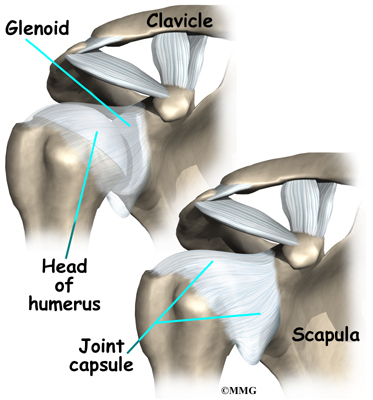
The most commonly affected tendons in the shoulder are the four rotator cuff tendons and one of the biceps tendons. The rotator cuff is made up of four small muscles and their tendons that cover the head of your upper arm bone and keep it in the shoulder socket. Your rotator cuff helps provide shoulder motion and stability.


What is Instability?

Instability:

Shoulder instability occurs when the head of the upper arm bone is forced out of the shoulder socket. This can happen as a result of a sudden injury or from overuse.

Shoulder dislocations can be partial, with the ball of the upper arm coming just partially out of the socket. This is called a subluxation. A complete dislocation means the ball comes all the way out of the socket.

Once the ligaments, tendons, and muscles around the shoulder become loose or torn, dislocations can occur repeatedly. Recurring dislocations, which may be partial or complete, cause pain and unsteadiness when you raise your arm or move it away from your body.

Repeated episodes of subluxations or dislocations lead to an increased risk of developing arthritis in the joint.


Friday, December 19, 2014

What is Impingement?

Impingement:

Shoulder impingement occurs when the top of the shoulder blade (acromion) puts pressure on the underlying soft tissues when the arm is lifted away from the body. As the arm is lifted, the acromion rubs, or "impinges" on, the rotator cuff tendons and bursa. This can lead to bursitis and tendinitis, causing pain and limiting movement. Over time, severe impingement can even lead to a rotator cuff tear.


Wednesday, December 17, 2014

What is Arthritis?

Arthritis:

Shoulder pain can also result from arthritis. There are many types of arthritis. The most common type of arthritis in the shoulder is osteoarthritis, also known as "wear and tear" arthritis. Symptoms, such as swelling, pain, and stiffness, typically begin during middle age. 

Osteoarthritis develops slowly and the pain it causes worsens over time.
Osteoarthritis, may be related to sports or work injuries and chronic wear and tear. Other types of arthritis can be related to rotator cuff tears, infection, or an inflammation of the joint lining.

Often people will avoid shoulder movements in an attempt to lessen arthritis pain. This sometimes leads to a tightening or stiffening of the soft tissue parts of the joint, resulting in a painful restriction of motion.


Tuesday, December 16, 2014

What is Bursitis?

Bursitis:

Bursae are small, fluid-filled sacs that are located in joints throughout the body, including the shoulder. They act as cushions between bones and the overlying soft tissues, and help reduce friction between the gliding muscles and the bone.

Sometimes, excessive use of the shoulder leads to inflammation and swelling of the bursa between the rotator cuff and part of the shoulder blade known as the acromion. The result is a condition known as subacromial bursitis. Bursitis often occurs in association with rotator cuff tendinitis. The many tissues in the shoulder can become inflamed and painful. Many daily activities, such as combing your hair or getting dressed, may become difficult.


Friday, November 21, 2014

Plantar Fasciitis

Plantar Fasciitis:

Who are risky?

1.    Gout
2.    Ankylosing Spondilitis
3.    Ruter’s Deases

C/F:

1.    Pain and tenderness of heel

Investigation:

1.    X-ray
2.    Serum Uric acid test
3.    HLA –B27

Treatment:

1.    Rest
2.    NSAIDs
3. Physiotherapy

(It takes may be 6 to 12 months to recover)


Thursday, November 20, 2014

De-Quervians Syndrome

De-Quervians Syndrome:

Who are risky?

1.    Middle age woman
2.    During pregnancy


Pathology:

1.    Thickening of tendon sheath.


C/F:

1.    Pain and swelling on styloid process of radious.
2.    Movement restricted.


Sign:

1.    Tenderness of styloid process.


Test:

1.    Finkelstrains test.


D/D:

1.    Arthritis at the base of the thumb.
2.    Scaphoid non-union.


Treatment:

1.    Rest
2.    NNSAID
3.    Steroid inj
4. Physiotherapy

Surgical Treatment:


1.    Division of the tendon sheath.

Friday, November 14, 2014

What Is Back Pain?

What Is Back Pain?

The spine is made up of 4 regions, the cervical (neck), thoracic (mid back), lumbar (low back) and sacral (base of spine). The bones that make up the spinal column are called vertebrae, with discs in between. Ligaments, muscles and tendons surround the spine to provide support and stability, and to control movement. So back pain problems can be complex.

How We Can Help?
We (Physiotherapist) assess and treat large number of back pain patients every day and use the latest treatment techniques to help our patients, including:

  • Joint Mobilizations
  • Manipulative Physiotherapy
  • Traction
  • McKenzie Therapy
  • Maitland Techniques
  • Specific Core Stabilization Exercises
  • Electrical Modalities
  • Thermal Modalities
  • Postural Advice

  • Acupuncture
  • Dry Needling
  • Massage
  • Exercise Techniques & Stretches
  • Ergonomic Advice
  • Lifting Techniques
  • Self-Management
  • Preventive Advice 

Thursday, November 13, 2014

How Neck Pain Can Be Treated By Exercise

How Neck Pain Can Be Treated By Exercise:
Neck Pain
We have all experienced neck pain to some degree- it may be brought on from sitting at a computer the whole day, from playing sports or possibly from an accident. Neck pain is a relatively common complaint, affecting up to 70% of individuals at some point during their life. Approximately 40-50% of the population suffers neck pain in any one year. Our Chartered Physiotherapist Niamh Connolly discusses the problem and some measures by which to reduce the chance of the problem occurring.

It tends to be a persistent and recurrent disorder where approximately 60% of individuals can expect to get some degree of on-going pain following their first episode.

The aim of physiotherapy rehabilitation of neck pain is to prevent a first episode from turning into chronic (>3 months) or recurrent pain.

The Neuromuscular system is one of the main reasons why people get a re-occurrence of their neck pain. The neuromuscular system is composed of nerves and muscles, these allow for innervations and movement of the muscles in the body. When a person undergoes pain and injury the strategies to control movement, posture and stability are compromised.

If neuromuscular function is altered this can cause:
·         Delayed activation of neck muscle
·         Changes in muscle size
·         Changes in muscle composition
·         Impaired postural endurance
·         Altered muscle activation movement

All of which can lead to neck pain:

Some examples of this type of altered function would be:

A reduced ability to maintain upright posture during a computer task, this reflects a low level of endurance in the muscles required to control the postural function of the spine, and over time if this is a repetitive position eg office / desk related job this may lead to neck/ shoulder pain.
How Is Neck Pain Treated?
The key principles in treating neck pain involve

·         Selectivity and specificity of exercise
·         Early rehabilitation
·         Pain-free rehabilitation
·         Rehabilitation for prevention of reoccurrence
How Can Exercises Help?
Exercise has been shown to improve neuromuscular impairments in people with neck pain however the type of exercise selected should be based on careful and precise physiotherapy assessment of these neuromuscular changes and therefore be specific to the impairments of the presenting patient.

This type of exercise usually commences early in the rehabilitation process and is used in combination with ‘hands on’/manual therapy if required; these exercises do not provoke pain and are designed to address the specific changes that have been identified via assessment in the muscle and neuromuscular system.
Exercises Used
The types of exercises used
·         target and activate the deep cervical muscles
·         retraining the endurance capacity of deep neck muscles
·         retrain the patterns of activation of the deep and superficial neck muscles
·         re-educate the use of muscles in posture and in functional tasks
·         address the strength and endurance  for functional requirements

Education and explanation regarding the rationale behind the treatment approach are a large component of this physiotherapy treatment as the patient’s compliance and contribution to the exercise program is critical.


Wednesday, November 12, 2014

Treating Lower Back Pain through the McKenzie Method:

Treating Lower Back Pain through the McKenzie Method:
We often discuss our physiotherapy treatments in our blog posts but do not always go into detail about the techniques and methods which are used by our physiotherapists. So today Mark Dockery of our Navan Road clinic in Dublin highlights how lower back pain is treated through the McKenzie method.
So What Is The McKenzie Method?

The McKenzie Method is a system of exercises used to elucidate the type of spinal issue a patient has and how best to treat it. It is commonly used worldwide in the diagnosis and treatment of low back pain, neck pain and peripheral joint complaints.
The method was created by New Zealand-based Physiotherapist Robin McKenzie (who passed away in May) in the early 1960s and is still in use today. The McKenzie Method is best applied with the aid of a Physiotherapist trained in the Method, who can diagnosis your particular problem and teach you the appropriate exercises to use at home.
The McKenzie Method implements primarily self-treatment strategies, and minimises manual therapy procedures, with the McKenzie-trained therapist supporting the patient with passive procedures only if an individual self-treatment programme is not fully effective. McKenzie himself states that self-treatment is the best way to achieve a lasting improvement of any type of back pain .
The 3 Focus Points Of The McKenzie Method
There are three main areas addressed by the McKenzie method for back and neck pain. They are posture, dysfunction and derangement . 
- Posture: End-range stress of normal structures.
- Dysfunction: End-range stress of shortened structures (i.e. scar tissue; fibrosis; nerve root adherence).
- Derangement: Anatomical disruption or displacement within the motion segment.
(The three mechanical syndromes – posture, dysfunction, and derangement – occur in all areas of the vertebral column, from the neck to the base of the back).
So What Might A Physiotherapist Advise?
Each distinct syndrome is addressed according to its unique nature, with mechanical procedures utilizing specific movement and positions. Examples of exercises prescribed by a Physiotherapist for a home-based programme might include:
- Lying prone (on your front). [McKenzie method treatment]
- Prone back extension (arching your back whilst lying on your front).
- Rotation mobilisation in extension (whilst lying prone).
- Standing posture exercises (importantly extension of the spinal column).
- Standing toe-touch.
- Pelvic side-shift movements (lateral tilt).
The McKenzie method encourages education and patient involvement, in managing their own treatment plans. This helps to reduce pain and restore normal function. This also may reduce the number of visits to the clinic. Overall most patients are able to treat themselves successfully when they are provided with the necessary information by their physiotherapist. Customised self treatment programmes will be provided by our physiotherapists should they deem this the correct method of treatment for you.


Thursday, November 6, 2014

De Quervain’s Disease:

De Quervain’s Disease:

Symptoms/Chief complain (C/C):

1.    Pain in the radial side of the dorsum of wrist.
2. Active movement of thumb is painful.

Signs/On Examination (O/E)

1.    Swelling may present on radial styloid.
2. Tenderness present on radial styloid.
3. Crepitus may present on palpation.
4. Abduction and extension of thumb against resistance is painful.

Treatment:

1. Complete rest of the hand with resting splint.
2. Ice.
3. UST with Naproxen gel.
4. NSAIDs.